Can 3D Printing Improve Your Medical Care?
So, you’re worried about getting, let’s say, a knee replacement. Will it be difficult? Will it hurt?
These are good questions. Every patient’s body is unique, and even the best surgeons do a bit of educated guessing to try to place a new knee as perfectly as possible. If the placement is even a bit off, there could be extra pain and stiffness, and it could take longer to recover.
But, what if, before your operation, your surgeon could make an exact replica of the parts of your knee—that’s your knee, as measured by your MRI and CT scan? Then, what if the surgeon could use it to plan the operation in advance and ensure precise placement of the new parts, which in turn means you’d have less pain, a speedy recovery, and a new knee that is just as good as the original? Better, actually.
That future is here. Some Yale Medicine surgeons now routinely use 3D printing (essentially producing a solid, three-dimensional object from a virtual digital model) to plan surgeries, design tools specific to an upcoming surgery and that particular patient’s anatomy, and even to print some of the parts used to replace defective ones in the body.
Daniel Wiznia, MD, a Yale Medicine orthopedic surgeon, is seeing significant improvements in his knee replacement patients since he started using 3D printing to plan their operations. He is one of several doctors who make regular use of a 3D printer at the Yale School of Engineering & Applied Science, a MakerBot machine that looks something like a microwave oven.
Vincent Wilczynski, deputy dean of the school and director of the Yale Center for Engineering Innovation & Design, remembers one of the first doctors who visited the lab several years ago, who used a printer to make a model of a 20-year-old patient’s knee tumor. “He printed the tumor every time the patient came in to get scanned, so the patient could hold the 3D model in his hands and feel how it was shrinking,” Wilczynski says.
There is more to come. Researchers are exploring ways to use 3D printing to make patient-specific transplant organs and eyes capable of translating light into electrical signals. While practical use of these things is still years away, researchers recently printed the world’s first 3D vascularized, engineered heart, using a patient’s own cells and biological materials.
Here are a few ways some Yale Medicine doctors are using 3D printing in patient care right now.
Easier, speedier knee replacement recovery
If you are having a knee replacement, 3D printing can make your surgery smoother, lessen the pain (which can be significant after knee surgery), and contribute to a quicker knee replacement recovery, Dr. Wiznia says. With this technology, your new knee can be implanted more accurately, leading to better biomechanics so the knee feels more natural when you move, with less wear on the implant, he adds.
“It’s a matter of precision,” Dr. Wiznia says, explaining that it lowers the risk of error compared to the traditional placement of a new knee. Following a 3D-printed model with cutting guides allows the surgeon to conduct surgery without significant positioning errors, he says.
When he is planning a surgery, Dr. Wiznia feeds data from the patient’s MRI into a computer program, allowing him to use software to plan where he wants to place the new knee, how he wants it to fit on the bone, the sizes of the implants and how the knee should bend kinematically. Then, he converts the information into a format that can be fed into a 3D printer so it will custom print surgical instruments. “The beautiful thing is, you don’t have to cut as much when you do the operation,” he says. “I use smaller incisions, because I’ve already modeled how to position the implants. There is less stretching of the ligaments and soft tissue, so there is less pain, blood loss, and length of stay in the hospital.”
He predicts knees implanted this way will have a longer life span. He also says pre-planning with 3D printing allows him to shave as much as 20 to 25 minutes off the time it takes to perform a knee operation. For the patient, this means less anesthesia, fewer blood transfusions, and less tourniquet time, he says.
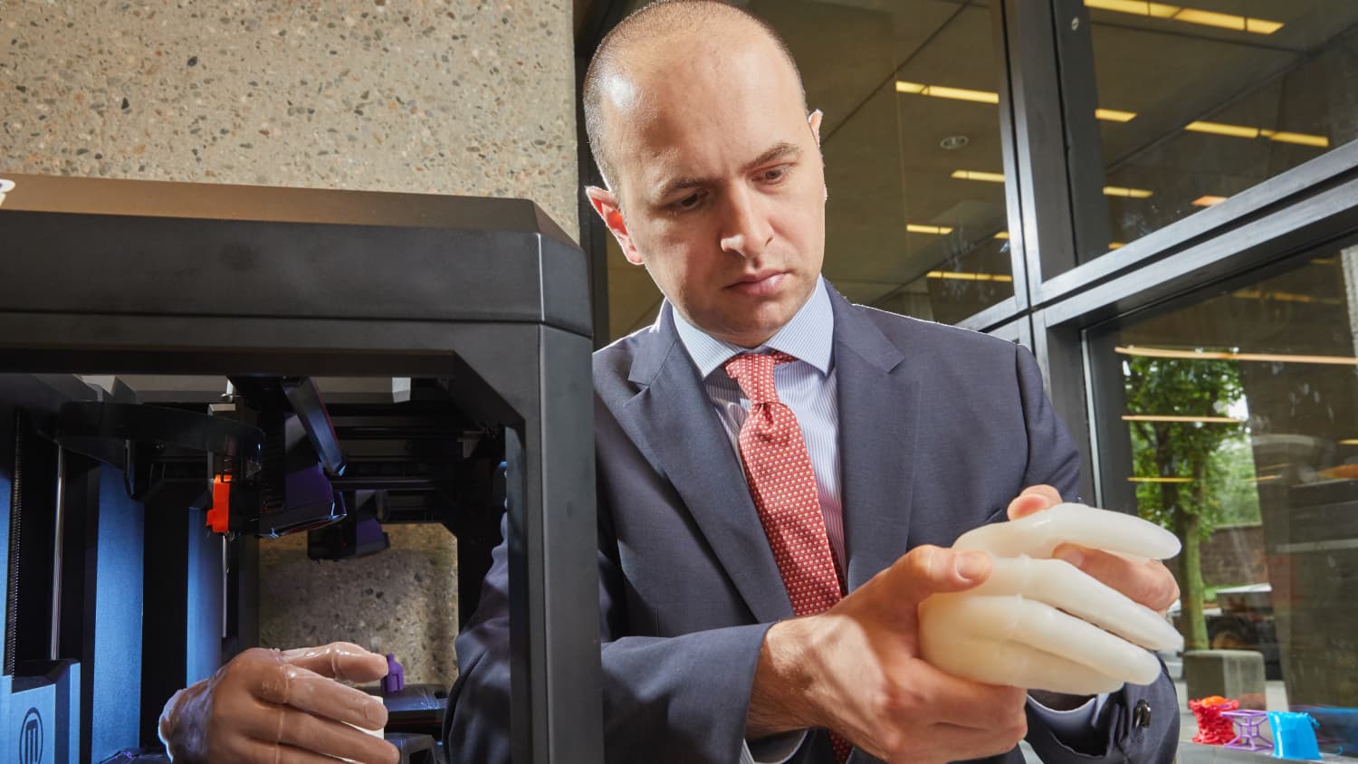
Planning operations with life-sized hand models
You slip on an ice patch and fall on your wrist, or you get mad and punch a wall. You can break a bone in your hand that way. Adnan Prsic, MD, a hand surgeon, decided it would be easier to fix this type of injury by first printing out a life-size model of your hand to better see what he and his residents will be working with. He has done this so many times he has amassed a collection of printed hands he keeps in a box in his office. Made of silicone, some are complete with "bones" inside made of varying types of plastics. If you bend a knuckle in one of these hands, you’ll hear a soft click (much like the legs in an old Barbie doll body). Dr. Prsic might “break” a bone inside the hand to simulate an injury.
Dr. Prsic is using 3D-printed hands in two ways. One is to help surgeons practice an especially complex hand surgery so they can better anticipate challenges. The second is to teach medical residents their craft—hand surgeons operate on small areas, so precision is key to sparing bone. He is collaborating on this with plastic surgeon Albert Woo, MD, from the Alpert Medical School at Brown University.
Other hand specialists have tried similar things with high-end 3D printers, but Dr. Prsic wants the capability to be available to anyone. So, he is making hands on the inexpensive MakerBot machine; he says each costs about $30, compared to $150 to make a hand on a more sophisticated machine. Inspired to become a hand surgeon in his native Bosnia, where he saw people come out of war with terrible hand injuries, Dr. Prsic says one of his future goals is to provide a model for inexpensive prosthetic hands that will be affordable for people in developing countries. He and Yale Medicine colleague James Clune, MD, are working on this together.
Easing anxiety when a birth defect is diagnosed
The more you can understand about a birth defect while the baby is in utero, the better you will be prepared to treat it, says Mert Ozan Bahtiyar, MD, director of the Yale Medicine Fetal Care Center. While it’s not currently possible to print out a model of a problem deep inside the body, like a heart defect, he envisions using 3D models of such problems as cleft palates and hand deformities. He believes these could be helpful psychologically to expectant parents.
He also has printed a placenta to show prospective mothers who he treats with twin-to-twin transfusion syndrome, a condition characterized by an imbalance in blood flow due to placental vessel connections. “I show them what the surface of the placenta looks like and talk about how the procedure is done."
Producing these 3D parts for expectant parents is more difficult than creating models of hands or hips. Doctors use ultrasounds when they perform imaging for pregnant women because it does not harm the unborn baby. But software to convert ultrasound to a 3D-printable file format isn’t as readily available as it is for CT scans. So, Dr. Bahtiyar is working with Xenophon Papademetris, PhD, professor of Radiology & Biomedical Imaging and Biomedical Engineering at Yale School of Medicine, and Praneeth Sadda, a Yale medical student, to develop what he needs. His ultimate goal is to keep a 3D printer in his office so he can print out pictures during patient visits.
“When I diagnose a fetal anomaly, the next step is counseling parents about how it will look,” says Dr. Bahtiyar, explaining that a 3D print is more powerful than an ultrasound, which provides a picture in two-dimensional black and white, with fields of gray. “I don’t think it will change the way we treat these problems. But pregnancy is not only the physical part, it is emotional as well, so maybe this will help a patient’s psyche. Maybe if they touch it, feel like it’s there, it makes them less anxious. It takes away the surprise.”
Restoring or adjusting to fix a face
Sometimes plastic surgeons are called upon to reposition a nose or create a stronger jaw, either for cosmetic purposes or to improve function. Besides repositioning parts of the face, their work may include facial reconstruction surgery and bone manipulation. Derek Steinbacher, DMD, MD, plastic surgeon and director of craniofacial medicine for Yale Medicine, has a varied collection of skulls, faces, and cutting guides, along with other 3D prints. A leading expert in the use of 3D imaging and printing in plastic surgery, he uses these techniques to aid in his treatment of complex cases such as rebuilding sections of the face or skull, treating congenital skull abnormalities, and nasal, orthognathic, and facial aesthetics.
He has been using 3D printing routinely since he arrived at Yale almost nine years ago, and has used it for almost 2,000 cases. He says it has revolutionized these surgeries, taking the process from “eyeballing intraoperatively” to a high level of planned precision that makes his work far more efficient, reproducible, and accurate.
The first step in bony 3D planning and printing is for the patient to have a CT scan of the area where he will operate. “That is converted to a virtual 3D image that can be manipulated—like an architect who builds something by first working with a 3D model in digital space,” Dr. Steinbacher explains. “In our case, we analyze and manipulate the 3D anatomy and gain insight into the patient’s diagnosis, walk through possible reconstructive scenarios, choose the final plan, and then print models and guides.”
Rhinoplasty (plastic surgery on the nose) is one operation where the approach is especially useful. “We look at a patient’s pre-existing nose, and we can see whether it is pointing left or right, or if it is somehow asymmetric. Then we manipulate it on the computer. There is some aesthetic and artistic preference that goes into it,” he says. “I also have to consider function.”
Dr. Steinbacher says, “it is possible from the 3D image to print a model of their nose or face with the actual expected post-op results.” He works closely with companies that specialize in this type of 3D printing. He has also printed actual parts to use as permanent implants in a new face, and plates that can be used to help move a jaw. Or, “if there is a tumor in the lower jaw, you can see exactly where that tumor is,” he says. He compares that with the opposite side of the face to see how he will reconstruct it once the tumor is removed.
Making complex operations for kids safer
A 3D model has prompted David Frumberg, MD, a pediatric orthopedic surgeon, to completely change the direction of a surgery that he has planned. Dr. Frumberg performs complicated surgeries for young children with such problems as limb and spinal deformities, as well as those with a variety of congenital or acquired conditions. He has used X-rays, CT scans, and MRI imaging to assess deformities and injuries and to plan an operation ahead of time, but he says 3D prints provide the most useful information.
“A life-size model of the actual bones that I can hold in my hand—there is something about it that changes everything,” he says. When he is planning a foot surgery, he is able to orient the model foot in different directions. “I can see what would happen if I shift one thing—or cut it one way as opposed to another during the surgery.”
Using a 3D-printed model was especially helpful when Dr. Frumberg performed foot and ankle surgery on a young girl from outside the U.S. with a rare congenital disorder that caused extreme abnormalities in her limb alignment. The girl did not have access to proper medical care as a child, and because doctors normally would have treated the issues when she was a toddler, there was no clear roadmap, he says. “If you are doing a surgery at this level of complexity, and you really need to know where all the bones are and maybe even practice, there is no better way.”
Dr. Frumberg says the models also seem to help children and families feel better about their procedures. When he was preparing to do a complicated operation on another girl’s foot, he showed her a 3D model he had printed, and drew on it with a Sharpie to show the various cuts he would make. “She was excited for the surgery after we did that,” Dr. Frumberg said. Later, he said, one of his medical students made a small 3D print of the model into a keychain she could keep.

