Multiple Sclerosis: How Scientists Developed a Breakthrough Drug
Imagine waking up to a world that blurs in front of you. Heading to the kitchen for breakfast, you’re hit with a wave of exhaustion, and your arms and legs feel like lead. This is the reality for the more than 400,000 people living with multiple sclerosis (MS), one of the most common neurological conditions in the United States. MS causes progressive nerve and brain damage as the body attacks the protective covering around each of its own nerve cells (called the myelin sheath). It causes vision loss, weakness and numbness in legs, and eventual disability. There are two types: relapsing-remitting and primary-progressive. In relapsing-remitting MS (the more common form), patients experience episodes of symptoms, followed by periods of recovery. In primary-progressive MS, there are no periods of relapse and the patient’s condition steadily declines.
While records of this condition date back hundreds of years, it is only in the past 25 years that the first drugs with modest efficacy were approved to treat MS. Still, there hasn’t really been a good way to control it—until last year. In 2017, the Food and Drug Administration (FDA) approved ocrelizumab, a medication that has been shown to significantly slow down the progression of the disease, and the first approved to treat primary-progressive MS. It was hailed in headlines as a breakthrough drug.
But why didn’t this drug come sooner?
Contrary to popular belief, most drug discoveries are not the product of one eureka! moment, but the result of decades of research, trial and error, and persistence woven together by scientists and doctors from around the world, each learning from others’ findings and mistakes.
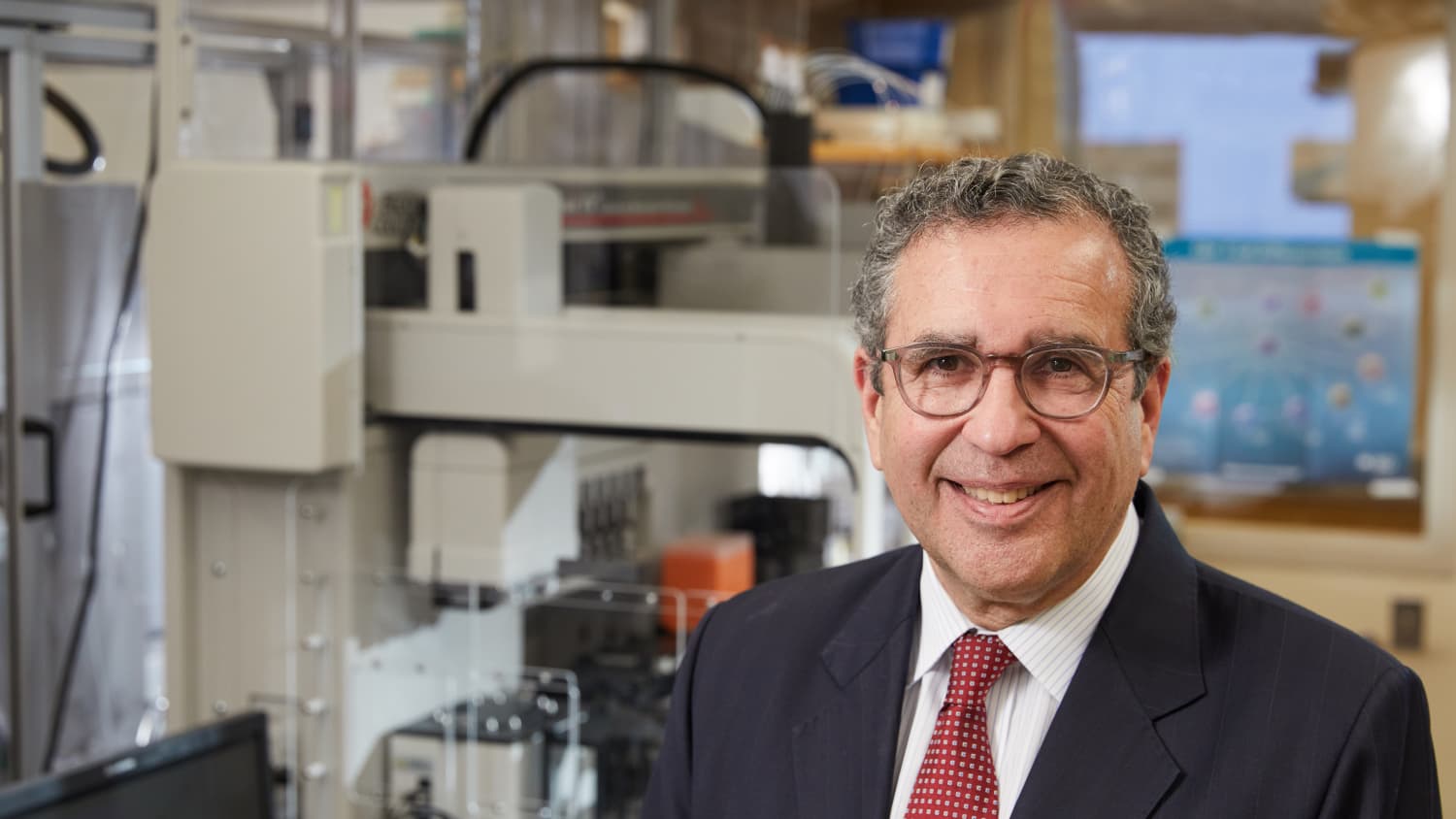
“Drug discovery can be a random walk,” says David A. Hafler, MD, chair of neurology at Yale Medicine. “There are myriad experiments a researcher could choose to do. The one that ultimately leads to the cure can be based largely on chance.”
Dr. Hafler would know: With 402 published papers, he is one of the most widely cited living neurologists and has decades of first-hand experience with the complexity of medicine. He also played a major role in identifying the biological processes underlying multiple sclerosis. “Ocrelizumab is a significant milestone in treating MS,” says Dr. Hafler. “What’s exciting about this drug is that it gives us insight into its cause.”
Here’s how it all came together.
Multiple sclerosis is a mysterious disease
In 1868, Jean-Marie Charcot, known as the “father of modern neurology,” dissected the brain of a deceased patient with (what we now think of as) multiple sclerosis and found hardened patches, indicating nerve damage, scattered throughout the white matter of the spinal cord, brain stem and brain. That was when the scientific community began to suspect that the condition, which had baffled physicians because of its constellation of symptoms with no apparent root cause, was a disorder affecting the nervous system. “It was also the first time anyone identified multiple sclerosis as a distinct disease,” says Dr. Hafler, who has a bust of Dr. Charcot sitting in his office.
Dr. Charcot’s findings were later corroborated when new microscope technology allowed scientists to observe nerve cells in more detail. In the early 1900s, James Dawson, a scientist at The University of Edinburgh, used this technology to see inflammation around blood vessels and damage to myelin (the fatty covering that protects nerve cells) of patients with multiple sclerosis. This damage was unlike anything doctors had seen before. So, what was causing it?
Because Dr. Dawson (and later researchers) had described inflammation around blood vessels, some scientists believed that MS was caused by a toxin circulating in the bloodstream that leaked into the brain. Others were able to mimic damage to myelin by blocking blood flow to the brain in lab animals, pointing to circulation problems as a potential cause. Still others thought that MS might be caused by a virus.
So, there were different theories, but a promising one emerged in 1933 when Thomas Rivers of the Rockefeller Institute in New York City injected Rhesus monkeys with virus-free myelin that, surprisingly, caused the immune system to attack its own body. Those monkeys then exhibited symptoms similar to patients with MS. This model, now called experimental autoimmune encephalomyelitis (EAE), was heralded as a breakthrough—it allowed scientists to look more closely at what was going on inside a living organism that showed the same type of nerve damage as patients with MS.
The model also suggested that MS was not caused by a virus. But just like in a viral reaction, the disease still seemed to involve the immune system. In 1960, neuroimmunologist Philip Paterson, in a now “classic” experiment, was able to transfer MS-like symptoms to healthy rats from EAE mice by injecting them with lymphocytes (a type of white blood cell involved in the immune response) from the affected animal. He had chosen lymphocytes because they were found in the plaque of the brains of deceased MS patients.
“By this point, attempts to prove the other hypotheses for MS were running into dead ends, while the immune system theory seemed to have legs,” says Dr. Hafler. “So, that’s the one we decided to pursue.”
MS turns the body against itself

Dr. Hafler was always fascinated by immunology and blood. He prepared a slide of his own blood and performed photomicroscopy using a toy microscope in fifth grade. The photograph was discovered this year by his mother Ann.
The immune system piqued Dr. Hafler’s interest—even as a child. As a 10-year-old, he started looking at his own red and white blood cells (after using a pin to prick himself) under a toy microscope. By sixth grade, he knew he wanted to be an immunologist. He later went on to study under several world-renowned scientists who researched MS, including Dale McFarlin, Henry Kunkel and Howard Weiner.
After graduating from medical school, Dr. Hafler wanted to tackle the unresolved questions surrounding multiple sclerosis. “I was interested in the brain and the immune system, and the two naturally came together,” he says.
By this point, studies using the EAE model had shown a connection between T cells (a white blood cell that’s part of the immune response) and multiple sclerosis. Dr. Hafler wanted to better understand these cells. In the 1990s, his lab was the first to recreate T cells that would attack the myelin protein and, in 2002, discovered a special type of T cell in humans that suppressed or calmed the immune system after it had done its job. These cells, called regulatory T cells, prevent the body from attacking its own cells in an autoimmune reaction. They were defective in patients with MS.
A genetic component
Around the same time, studies began to emerge showing that twins with MS had a related risk of developing the condition. But it was complicated: The research showed that MS occurred in a small percentage of siblings and nonidentical twins but in about 25 percent of identical twins. This meant that genetics had something to do MS, but it wasn’t the sole cause.
This genetic component was intriguing to Dr. Hafler. “I was getting frustrated with immunology—there were too many moving pieces, and it was hard to know which part of the immune system to study,” he says. But with genetics, there was a clear path forward.
Dr. Hafler helped found the International Multiple Sclerosis Genetics consortium, which brought together researchers from 30 institutions in 18 countries. By combining data from multiple studies, the consortium assembled genomic data from thousands of people diagnosed with MS and compared them with thousands of people without the condition. As a result, they were able to identify 233 variants of genes that, combined together, increased a person’s chance of developing MS.
An accidental discovery
The established animal model scientists were using to study MS still didn’t mimic the condition completely. This was in Stephen Hauser’s mind while he was working in his lab in the early 1990s. The scientist had remembered something his mentor had told him while he was completing his neurology residency at Massachusetts General Hospital: The pathologies of the EAE model and multiple sclerosis were different—the tissue changes in the mice with MS-like symptoms didn’t completely match those in human MS.
Maybe the reason why his colleagues hadn’t yet been able to figure out the cause of MS was because they had been performing experiments on an inaccurate model.
Dr. Hauser spent years experimenting with various models, and in 1995, his research team was finally able to induce MS that looked exactly like the human version. But there was something more: The transfer of T cells didn’t cause MS like it had in the EAE model. It was only after they transferred both T cells and B-cell antibodies together that they were able to induce multiple sclerosis.
This was a surprise. For decades, scientists had focused only on T cells. So how did B cells—another type of cell in the immune system that’s part of the immune response—fit into this picture?
Dr. Hauser’s hypothesis was that B cells were somehow triggering T cells to cause an MS attack. He wanted to try testing medications that would target B cells. Coincidentally, a new drug called rituximab had just been approved by the FDA to treat the autoimmune disease called non-Hodgkin’s lymphoma. It was the first approved treatment directed against B cells.
Dr. Hauser’s team published a groundbreaking study in 2008. The study followed 104 patients with MS, 69 of whom had been treated with rituximab. The formation of new brain lesions (a common marker of attacks to the myelin in MS patients) in rituximab patients were reduced by 91 percent compared to those on the placebo. The study showed not only that B cells somehow played a role in causing the disease, but that when B cells were depleted by the drug, brain inflammation was almost immediately shut down.
Six years later, after multiple iterations of clinical trials, the FDA approved a medication closely modeled after rituximab. That medication was ocrelizumab.
Although it’s not clear how the drug works, scientists know that it deletes circulating B cells and with that, prevents the abnormal T cell immune response that attacks the myelin.
Ocrelizumab is the first drug approved for primary-progressive MS, the most aggressive form of the disease, in which the brain and body steadily deteriorate. Clinical trials showed that patients taking doses of ocrelizumab showed significantly fewer brain lesions after 24 weeks, and a lower relapse rate for patients with relapsing-remitting MS (the most common type of MS). The drug beat out interferon beta 1a, one of the most commonly used drugs to treat MS, in reducing annual relapse rate and the number of lesions seen on MRI scans.
Understanding MS is an ongoing journey
So, there are some answers. But more questions remain. What are B cells doing to the T cells to trigger MS? And what role do genes have in all of this?
These are questions that Dr. Hafler’s lab is wading through. Currently, they are using new technology called single cell RNA sequencing to observe what types of cells are affected before and after treatment in MS patients to better understand the basic biology underlying the condition. His research also represents a departure from the usual way of doing science.
“While most of science is about model-making, we have decided to observe first and create a hypothesis from that,” Dr. Hafler says.
In addition, previous research has shown that salt and fat may contribute to dysfunction in regulatory T cells, which suggests that environmental factors may be a piece of the puzzle. Dr. Hafler’s lab is continuing to investigate exactly how those factors might influence a patient’s risk for MS.
Ocrelizumab itself is not the end of the road either—there haven’t been studies to look at its long-term side effects. On the plus side, its clinical trial results outperformed every other MS drug available on the market and reduced the annual relapse rate for patients with relapsing forms of MS by 50 percent. However, it offers only about a 25 percent benefit to patients with a primary-progressive form of the disease. This leads researchers to wonder if there are different biological mechanisms at work.
Still, Dr. Hafler remains hopeful. In 2014, his lab published a paper in Nature that mapped the genes of cells involved in various conditions. They found that the cells most involved in passing down multiple sclerosis are immune cells, not brain cells, which further confirmed that multiple sclerosis is an autoimmune disease rather than a neurological one.
“It’s been a long and winding road to where we are now,” says Dr. Hafler. “And it will probably continue to be until we find all the answers. But we have the best scientists working on it, and I’m confident it will come together.”