Musculoskeletal Ultrasound
Overview
Ultrasound is a safe and painless way of using sound waves to look at parts of the human anatomy. The technology works on a principle similar to that used in sonar, which helps submarines navigate, and echolocation, which allows bats to see their surroundings even on moonless nights.
Ultrasound has many applications, from diagnosing injuries such as muscle tears and chronic conditions, including rheumatoid arthritis, to guiding caregivers through diagnostic and therapeutic procedures. No radiation or injectable contrast agent is needed.
"Musculoskeletal ultrasound allows physicians to see—in high resolution—a person’s muscles, tendons, ligaments, nerves and joints," says Risa H. Kent, MD, a Yale Medicine radiologist.
At Yale Medicine, our radiologists, clinicians and technicians are highly trained and experienced with our state-of-the-art technology, often allowing us to diagnose and treat a condition during the same ultrasound visit.
What makes musculoskeletal ultrasound unique?
In addition to the high-resolution images, the technology allows you to move around during the procedure. “We can focus on the exact location of the patient’s pain and use dynamic imaging to determine how motion affects the area of concern," says Dr. Kent.
Dynamic imaging creates clear pictures in real-time as you move your arm, leg, or other area of the body. “Using the ultrasound’s Doppler effect, which tracks moving substances,” she says, “we can also focus on arteries and veins—and determine how blood is flowing.”
What can musculoskeletal ultrasound diagnose?
Musculoskeletal ultrasound can help to diagnose a range of injuries and chronic conditions, including tendonitis, bursitis, carpal tunnel syndrome, rotator cuff tears, joint problems, and masses such as tumors or cysts.
How does musculoskeletal ultrasound work?
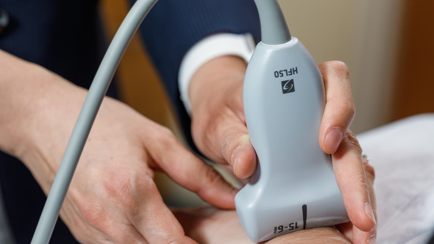
During a musculoskeletal ultrasound, a hand-held scanning device called a transducer is pressed on your skin. The transducer sends out inaudible, high-pitched sound waves that travel through the body.
Denser substances such as bone reflect the waves back while liquid, including water, allows the waves to pass through. The transducer converts wave activity into accurate pictures of muscle, tendon, and other structures.
What should patients expect during the procedure?
A radiologist, your orthopedist or rheumatologist, or an ultrasound technician may do the scanning. You will be asked to to sit or lie down, depending on where the pain, or other issue is situated.
A layer of gel is applied to the transducer, which helps it transmit and receive sound waves. Next, the gelled device is placed against your skin, near the affected area.
The professional administering the sonogram will slide the transducer on the skin, and may ask you to move the affected area to reproduce symptoms.
What are some benefits of the procedure?
Musculoskeletal ultrasound can often provide a clearer picture of your musculature, tendons and other affected structures than either an X-ray, CT or MRI—or the test may compliment those other procedures.
“By moving the transducer,” Dr. Kent says, “the professional performing the ultrasound can view relevant anatomy at different angles, taking still pictures as needed.” By administering the ultrasound there's a better understanding of what may be causing the issue. (Dr. Kent says that MRI is the preferred test to evaluate structures deep within a joint.)
Ultrasound doesn’t require getting an injection of a contrast agent. Because it doesn’t use radiation, it poses none of the health risks associated with X-rays. Unlike in the case of MRI, you aren't confined in an enclosed space, so you won’t experience claustrophobia.
Who is a candidate for musculoskeletal ultrasound?
Anyone experiencing arthritis or muscle pain, with or without inflammation, might get a referral for the procedure. Other candidates may include patients with soft tissue masses or infections or athletes or others who have experienced an acute injury or have a chronic condition.
Because musculoskeletal ultrasound poses minimal discomfort or risk, Dr. Kent emphasizes that “anyone of any age, young or old, pregnant or not, may be eligible for the procedure.”
What makes Yale Medicine’s approach to musculoskeletal ultrasound unique?
At Yale Medicine, radiologists and clinicians, in addition to technicians, are trained to become experts with the technology. Because of this, Dr. Kent says, there are situations where a person’s condition may be diagnosed and treated during the same ultrasound visit.
“It’s very gratifying—for both the caregiver and the person undergoing the procedure,” she says.