Melanocytic Nevi (Moles)
Overview
Melanocytic nevus is the medical term for a mole. Nevi can appear anywhere on the body. They are benign (non-cancerous) and typically do not require treatment. A very small percentage of melanocytic nevi may develop a melanoma within them. Of note, the majority of cutaneous melanomas arise within normally appearing skin.
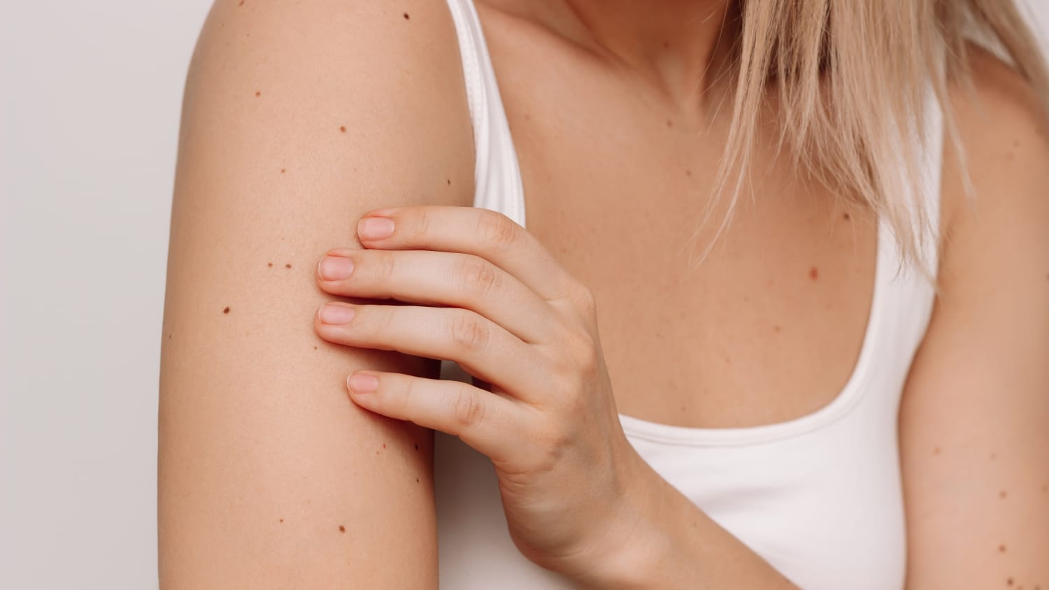
Benign nevi are usually a single color, ranging from skin-colored to dark brown. They are typically round or oval-shaped. In addition, benign moles are symmetric, that is, when a line is drawn within them, the two halves have the same appearance. Most melanocytic nevi are the size of a pencil eraser or smaller. When larger in size, they may be referred to as atypical nevi or they may represent moles present since birth (congenital nevi). About 1 in 100 babies are born with a congenital nevus.
“Nevi begin to appear during childhood and increase in number until early to mid-adulthood.
As nevi age, they can become elevated but also soft,” says Yale Medicine dermatologist Jean Bolognia, MD. “If a nevus becomes elevated and is firm to the touch, it should receive medical attention. When there is a suspicion of cutaneous melanoma, a biopsy will be performed.”
What are melanocytic nevi?
Melanocytic nevi are benign tumors that that arise in the skin. They have different sizes and colors as outlined above. Benign nevi are usually round or oval-shaped and are uniform in color. There are more nevi in areas of the body that have greater long-term exposure to the sun, such as the outer arm compared with the inner arm.
Cells within the skin called melanocytes produce a pigment named melanin. This pigment is what gives melanocytic nevi a tan to dark brown color. Melanin is also responsible for overall skin color and the darkening seen with a sun tan.
What leads to melanocytic nevi?
Melanocytic nevi are a reflection of genetic factors, such as family history, and environmental factors—primarily, sun exposure.
What are the clinical characteristics of melanocytic nevi?
- Tan to dark brown, pale pink, and occasionally black in color
- Round or oval in shape
- Smooth borders
- Uniform color throughout
- Symmetry (when a line is drawn within them, the two halves have the same appearance)
As melanocytic nevi age, they often become lighter in color. They may also elevate but should become softer to the touch.
What are atypical nevi?
Atypical nevi have a number of clinical features that overlap with cutaneous melanoma, but they are benign lesions. These features include:
- Asymmetry (when a line is drawn down the middle of the mole, the two sides are not the same)
- Irregular, rather than smooth, round borders
- Variation in color within the mole, with more than one color
- Larger than a pencil eraser
Patients with numerous (over 50, for instance) benign nevi and/or multiple atypical nevi are at increased risk of melanoma and should be followed by a dermatologist. Because the clinical features of atypical nevi and cutaneous melanoma overlap, biopsies of atypical nevi are performed when there is concern regarding possible melanoma.
How are melanocytic nevi managed?
Patients with a changing mole often seek medical attention. Sometimes, the explanation is irritation due to location while other times there are concerning features. In the latter situation, a biopsy is routinely performed. In addition, patients with a personal or family history of melanoma, numerous nevi, and/or atypical nevi should have total body skin examinations on a regular basis.
Dermatologists may use a hand-held device known as a dermatoscope to closely examine individual moles to assist in the clinical diagnosis. A dermatoscope magnifies the designated lesion, in addition to accentuating pigment patterns. Some dermatologists photograph certain nevi to monitor changes over time.
For the detection of cutaneous melanomas, there is an “ABCDE” rule, designed to assist in remembering the clinical features of melanoma. As noted above, these findings can overlap with those of atypical nevi. This rule consists of:
- Asymmetry
- Borders—irregular or blurred
- Color—multiple colors and areas of darkening
- Diameter—greater than 6 mm
- Evolving—changes in size, shape, or color; itching or bleeding
How are melanocytic nevi treated?
The majority of melanocytic nevi do not require treatment. Nevi that are benign but bothersome to the patient because of appearance or irritation can be removed. Moles are removed by the shave technique or suture surgery.
Atypical melanocytic nevi are biopsied when the clinician is concerned about the possibility of cutaneous melanoma.