Ocular Hypertension
Overview
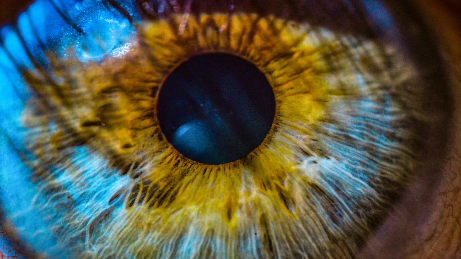
Ocular hypertension is a condition that occurs when pressure within the eye increases without affecting a person’s vision or damaging their eye anatomy. It arises when fluid doesn’t drain out of the eye at its regular rate, resulting in a buildup that increases intraocular (within the eye) pressure. Ocular hypertension may be an early sign of glaucoma, an eye disease that can cause irreversible vision loss over time if it is not treated.
Ocular hypertension does not cause any noticeable symptoms. A doctor uses special equipment to measure a patient’s intraocular pressure during an eye exam to assess their eye health. Intraocular pressure levels greater than 21 mm Hg confirm a diagnosis of ocular hypertension.
Older adults are more likely than those under age 40 to have ocular hypertension, which is more likely to develop as people age. An estimated 4% to 10% of Americans aged 40 and older have ocular hypertension. It’s possible to have the condition in one or both eyes.
Once a patient is diagnosed with ocular hypertension, doctors will assess their risk of developing glaucoma. This assessment helps doctors determine whether or not treatment is needed. When appropriate, doctors prescribe eye drops, which help to manage ocular hypertension and reduce the risk of glaucoma. In rare cases, surgery may be recommended to lower intraocular pressure.
Ocular hypertension is not caused by, or related to, hypertension (high blood pressure), although both conditions have similar-sounding names.
What is ocular hypertension?
Ocular hypertension is a term that refers to pressure within the eye that is higher than it should be. Although people with ocular hypertension don’t experience changes to their vision or eye anatomy, the condition can lead to glaucoma, which causes structural eye damage and vision loss.
Doctors monitor the eye health of patients with ocular hypertension because it may lead to glaucoma. However, an ocular hypertension diagnosis does not always develop into glaucoma.
All people have clear fluid, called aqueous humor, in the front portion of each eye, between the cornea (the clear covering at the front of the eye) and the lens (which focuses on objects, then creates images on the retina at the back of the eye). Aqueous humor, produced within the eye continuously, supplies the eye with nutrients and oxygen and removes waste products and debris. It also helps maintain the intraocular pressure and shape of the eye.
When a person has ocular hypertension, the aqueous humor doesn’t drain out of the eye as readily as it should, which leads to increased pressure within the eye. A blockage may develop within the eye, preventing the fluid from exiting through its usual routes.
What causes ocular hypertension?
Increased intraocular pressure may occur if too much fluid enters the eye or too little fluid drains from the eye.
Different conditions may prevent the aqueous humor from exiting the eye at the expected rate, including:
- Uveitis (inflammation of the middle portion of the eye)
- Pigment dispersion syndrome, in which tiny flecks of pigment from the iris (the colored portion of the eye) float around inside the eye, blocking the trabecular meshwork, a spongy tissue in the drainage angle. Aqueous humor passes out of the eye through the trabecular meshwork
- Pseudoexfoliation syndrome, when microscopic bits of protein fiber build up at various sites in the body, including the eye, where they may block fluid from draining out of the eye
- A tumor within the eye
- Large cataracts that block drainage routes
- Damage to the eye caused by injury or surgery
- Certain medications, including corticosteroids
- Chronic angle-closure, a condition in which the iris blocks the trabecular meshwork, thereby obstructing the flow of aqueous humor out of the eye
What are the symptoms of ocular hypertension?
Ocular hypertension doesn’t cause symptoms. People won’t know they have the condition unless they are diagnosed by an eye doctor during an office visit.
Because the symptoms are silent, people should see their eye doctors regularly so that the condition can be diagnosed early.
What are the risk factors for ocular hypertension?
People are at increased risk of ocular hypertension if they:
- Are age 40 or older
- Have a family history of ocular hypertension and/or glaucoma
- Have severe nearsightedness (myopia)
- Have severe farsightedness (hyperopia)
- Have diabetes
- Have high blood pressure
- Experienced an eye injury
- Have had eye surgery
- Have previously had eye injections
- Take, or have taken, steroid medications for long periods of time
- Have pigment dispersion syndrome
- Have pseudoexfoliation syndrome
- Use certain medications such as topiramate
How is ocular hypertension diagnosed?
Ophthalmologists (eye doctors who perform medical and surgical treatments for eye conditions) and optometrists typically diagnose ocular hypertension during routine eye exams. They can confirm the presence of the condition after asking a patient about their medical history, performing an examination, and offering diagnostic tests.
When speaking with an eye doctor about your overall health, be sure to mention if you have diabetes or high blood pressure. It’s also important to share details about any family history of glaucoma or ocular hypertension. If you have had eye surgery, eye injections, or a past eye injury, let the doctor know.
Eye doctors can’t tell that a patient has ocular hypertension just by looking at their eyes; they can discover the condition during a comprehensive eye exam, which includes different tests and assessments to check on a patient’s overall eye health.
- Tonometry. Your doctor will measure your eye pressure using a procedure called tonometry. There are several types of tonometry. In the most accurate type, known as applanation tonometry, the provider will numb the patient’s eyes with eyedrops and temporarily stain the eyes using a dye that may be given via eyedrops or a strip of paper with orange dye on it. With the patient’s chin resting on a slit lamp, the provider directs an applanation tonometer toward your eye until it touches the cornea. The device measures the how much pressure it takes to flatten part of the cornea, which allows the provider to determine the intraocular pressure. Another method known as noncontact tonometry involves the use of a special device to direct a puff of air at the eye to flatten the central part of the cornea. The device automatically calculates the intraocular pressure. Noncontact tonometry does not require numbing drops. Intraocular pressure between 12 mm Hg and 21 mm Hg is considered normal. Measurements higher than 21 mm Hg on two separate occasions are considered ocular hypertension.
- Goniscopy. If the patient’s intraocular pressure is elevated or if the provider suspects the patient may have other eye problems, additional tests may be required, including goniscopy. This test is performed while the patient places their chin and forehead against a slit lamp. The doctor places a special contact lens onto the surface of the patient’s eye. This contact lens has mirrors on it. The light that shines through the slit lamp is aimed at the drainage angle, where fluid normally exits the eye at the intersection of the iris and the sclera (the white part of the eye). Doctors can see whether fluid is flowing normally or if a blockage exists. If there’s a blockage, doctors can usually identify the cause.
Other tests may be also performed to determine whether there is vision loss or any damage to the optic nerve, or to measure the thickness of the cornea, which may impact the patient’s risk of developing glaucoma. If tests reveal vision loss or damage to the optic nerve, the provider will diagnose the patient with glaucoma.
How is ocular hypertension treated?
Ocular hypertension doesn’t always require treatment. Doctors determine whether a patient with the condition has a low, moderate, or high risk of developing glaucoma over the next five to 10 years. If a patient’s risk is moderate to high, doctors typically treat the condition (more on that below). Doctors are more likely to monitor ocular hypertension if the risk is low.
Doctors typically prescribe eye drops to lower a patient’s intraocular pressure by 20% to 25%, thereby reducing ocular hypertension. Medications may be prescribed by themselves or together. Options include:
- Prostaglandins
- Beta blockers
- Carbonic anhydrase inhibitors
- Alpha agonists
- Netarsudil, a rho-kinase inhibitor
In some cases, doctors recommend laser surgical procedures to lower intraocular pressure. However, most patients will not need such a procedure.
What is the outlook for people with ocular hypertension?
Ocular hypertension cannot be cured, but it can be managed with medication to reduce the risk of glaucoma.
People who receive treatment for ocular hypertension may be up to 50% less likely to develop glaucoma. Still, some people treated for ocular hypertension eventually develop glaucoma. This is less likely to happen among patients who see their eye doctor regularly and use the medication prescribed as directed.
This article was medically reviewed by Yale Medicine ophthalmologist David Silverstone, MD.