Glaucoma
Overview
If you have a family history or any other risk factor for glaucoma, you know how important it is to make regular visits to the eye doctor. That's because glaucoma is one of the primary causes of blindness in the United States.
“Glaucoma is actually a group of diseases with the common characteristic of optic nerve change that can result in vision loss,” says Ji Liu, MD, a Yale Medicine glaucoma specialist.
Yale Medicine's ophthalmologists have an international reputation for their work with glaucoma and other diseases of the eye. They use the latest tools to evaluate and manage people who are at risk and have been successful in preserving vision even for people with complicated cases of glaucoma.
What are the most common types of glaucoma?
There are two main types of glaucoma, both marked by an increase in intraocular pressure, which is pressure behind the eye. Normally, a nourishing fluid flows through the eye’s anterior chamber (an area in the front of the eye) and exits smoothly through an area of tissue called the trabecular meshwork to a canal-like drainage system. (The trabecular meshwork sits at the end of the chamber, where the iris and cornea meet to form an angle.)
When the fluid does not drain efficiently and pressure within the eye builds, it damages the optic nerve.
Open-angle glaucoma is the most common form of glaucoma in the United States. In open-angle glaucoma, the drainage angle is not visibly obstructed, but buildup of fluid and pressure causes damage. Open angle glaucoma can be primary, meaning that it does not have an identifiable cause; or secondary, meaning that it is caused by something else, such as inflammation, trauma or a response to steroids. “The exact mechanism is not fully understood,” says Dr. Liu. “However, it is important to lower the intraocular pressure to preserve vision.”
Narrow-angle glaucoma, a less-common form of the condition, occurs when the angle has decreased in size, limiting the flow of fluid through the drainage structure. Narrow angle can remain the same without becoming worse, but it can also progress to acute angle-closure glaucoma, a dangerous condition that develops very quickly and threatens vision.
Narrow angle can also lead to chronic angle-closure glaucoma with intermittent or persistent intraocular pressure elevation, and eventually optic nerve damage and visual function loss. (If there are no changes to vision or pressure elevation, the condition is called narrow angle glaucoma suspect.)
What are the risk factors for glaucoma?
Risk factors for glaucoma include confirmed elevated intraocular pressure (your doctor will look for a reading of pressure within the eye that is above 22 millimeters of mercury), a family history of glaucoma, age, and a thin cornea.
“Some data also suggest that diabetes and high or low blood pressure may be risk factors for developing glaucoma, but not all studies agree,” says Dr. Liu. People who have myopia (nearsightedness), obstructive sleep apnea or migraine headaches, or those who have experienced Raynaud’s phenomenon (abnormally reduced blood flow to fingers and toes in response to cold temperature) may also be at risk for developing glaucoma.
Although high intraocular pressure is a risk factor for developing glaucoma, not all glaucoma is related to high pressure. It is also true that some people with documented intraocular pressure above the normal range have no signs of optic nerve damage.
“We don’t treat everyone if they have an elevated pressure reading without any disturbance to their visual field or optic nerve,” Dr. Liu says. “But we closely monitor them because they have the potential to develop glaucoma.”
What are the symptoms of glaucoma?
Most types of glaucoma develop slowly with no symptoms at the early stages of disease.
“When glaucoma becomes moderate or severe, patients may notice blurred vision or missing parts of their visual field,” says Dr. Liu. “Some patients may not have any noticeable symptoms until the latest stages, which makes this disease particularly dangerous.”
In contrast, though, acute angle-closure glaucoma can develop within hours when the angle becomes blocked, preventing drainage and causing pressure in the eye to rise rapidly. Acute angle-closure glaucoma can cause eye pain, headache, decreased vision, and sometimes nausea and vomiting. If this condition is not treated immediately, vision may be permanently lost.
How is glaucoma diagnosed?
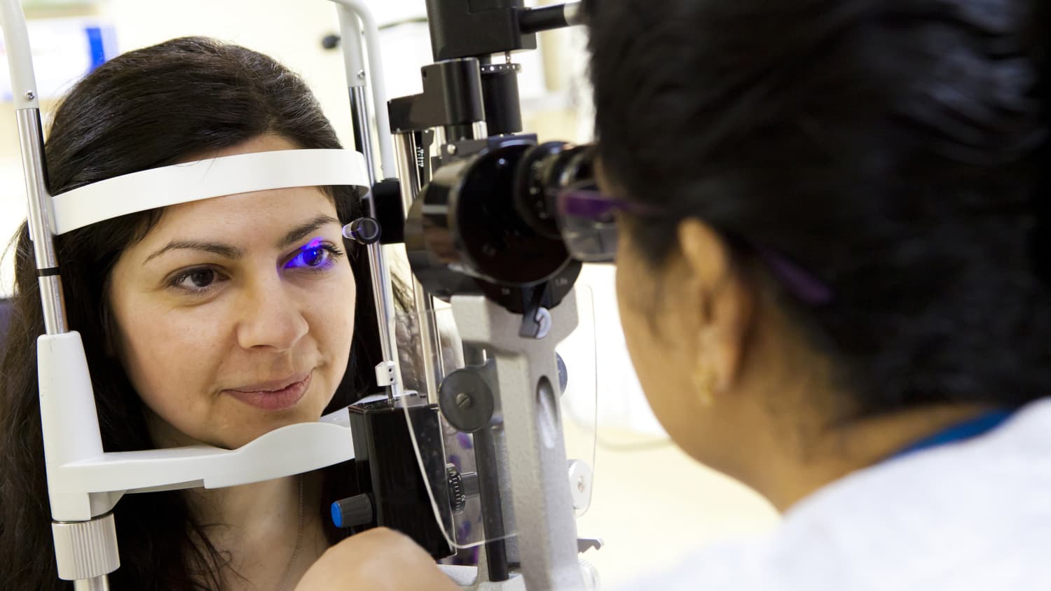
Glaucoma is diagnosed during a full eye exam. This includes an examination with a noninvasive slit-lamp (a microscope combined with a light source) to visualize the optic nerve and the angle, visual field testing to evaluate peripheral vision, and use of the Goldmann tonometer, which is the noninvasive tool that is considered to be the gold standard examination for measuring intraocular pressure.
Ophthalmologists at Yale Medicine often use optical coherence tomography, another noninvasive test that can provide high-resolution images of important structures of the eye.
“Some of these are performed as part of a routine eye examination, and some are performed on people who have glaucoma or those being monitored because they are suspected of having glaucoma,” says Dr. Liu. “But everyone over 40, or those who have a family history of glaucoma— including second-degree relatives, aunts, uncles and grandparents—should have a baseline exam that looks at the integrity and condition of the optic nerve.”
What are the treatment options for glaucoma?
“The only proven effective treatment for glaucoma is lowering intraocular pressure,” says Dr. Liu. Lower intraocular pressure can be achieved with the use of prescription eye drops, laser treatment or surgery. The recommended treatment is usually selected based on the severity of the glaucoma.
Early diagnosis and treatment of glaucoma are very important. “Treatment is initiated to stop the progression of the disease,” Dr. Liu says, “but it will not restore the vision that has been lost.”
Eye drops: Often the first line of treatment, they can either increase the outflow of fluid or decrease the production of fluid. Sometimes, patients may be prescribed multiple medications, some of which may require several doses throughout the day.
Laser: If eye drops don’t lower pressure enough to stop the progression of glaucoma, laser procedures may be recommended; they can also be used as a first line of treatment. Performed in a doctor’s office, laser procedures vary depending on the type of glaucoma, but they are painless and take just a few minutes to complete.
The most common laser treatment for open-angle glaucoma is laser trabeculoplasty, which reconstructs the trabecular meshwork to improve drainage. Laser peripheral iridotomy, which may be performed for narrow angle or angle-closure glaucoma, is used to make an opening in the iris of the eye, allowing fluid to bypass the normal route and reduce pressure within the eye.
Surgery: For more severe cases, or when medications and laser therapy do not improve intraocular pressure, surgery may be recommended. Minimally invasive glaucoma surgeries can help with fluid drainage and reduce the eye pressure. In people with advanced disease, a trabeculectomy that creates a small passageway so fluid can drain, or a drainage device (a shunt) that is permanently implanted in the eye, may be needed.
All of these operations are performed in outpatient settings.
“Compared with cataract surgery, these procedures carry higher risk,” Dr. Liu says. “However, if the intraocular pressure is not well controlled with medications or laser therapy, surgery may be the only remaining option to preserve vision.”
What makes Yale Medicine’s approach to glaucoma unique?
Every Yale Medicine patient is treated by a specialist who uses comprehensive eye exams, advanced imaging tests and state-of-the-art technology to fully evaluate and manage glaucoma and all eye conditions.
Using minimally invasive surgical techniques, sometimes performed in conjunction with cataract surgery, our ophthalmologists often have great success in preserving vision and halting the progression of glaucoma.