Mohs Surgery for Skin Cancer
Overview
Mohs micrographic surgery has been a game-changer in treating skin cancers such as basal cell carcinoma and squamous cell carcinoma.
In this procedure, the cancer is removed layer by layer, and the tissue is examined under a microscope after each step, so the dermatologist can confirm that all of the cancer cells have been eliminated. It maximizes the chances of removing all of the abnormal cells while still preserving as much of the normal skin tissue as possible.
Cure rates for skin cancer using Mohs surgery can reach up to 99%, and the chances of lasting, disfiguring scars are minimal. Yale Medicine dermatologic surgeons have specialized expertise in performing Mohs surgery.
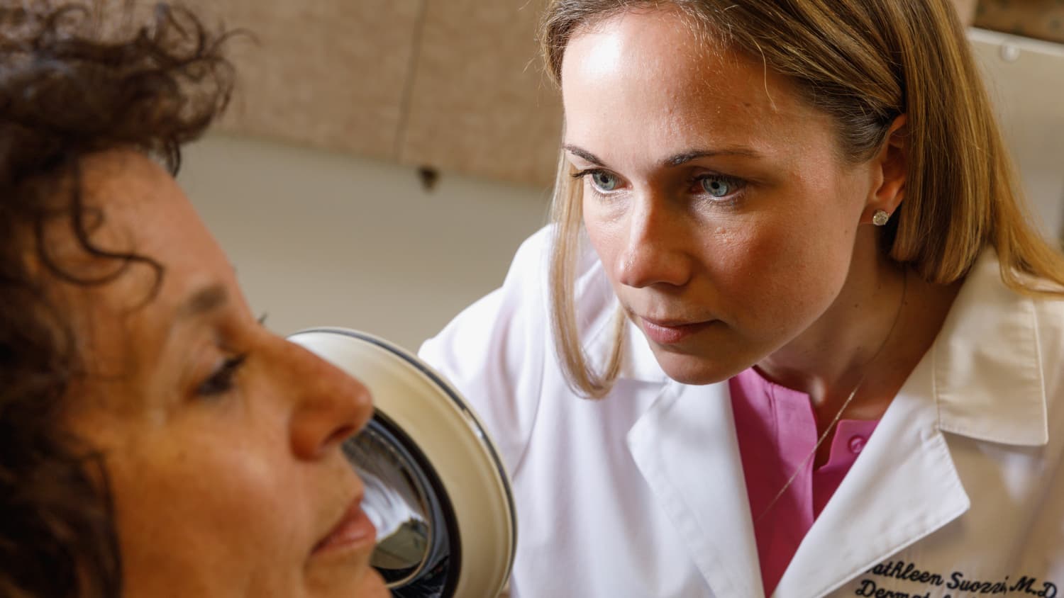
What is Mohs surgery?
“Mohs surgery is based on the notion that normal pathology specimens, cut like a bread loaf, evaluate only about 3 percent of the total surface area of the margins of the cancer,” says David J. Leffell, MD, a Yale Medicine dermatologist and section chief of Dermatologic Surgery. “By contrast, the Mohs technique allows evaluation of the complete surface area.”
This is important, Dr. Leffell says, because many basal cell cancers and squamous cell cancers grow with microscopic fingerlike projections or roots can be missed with traditional excision and examination of the tissue (another technique to treat basal and squamous cell carcinomas).
As part of the Mohs technique, the dermatologist not only removes the cancer from the skin but also “maps it out” with special colored inks. The dermatologist then evaluates it under a standard light microscope. Dermatologists must be specially trained to perform Mohs surgery, and it is performed in the doctor's office in just a few hours.
What skin conditions are best treated with the Mohs surgery?
There are some situations in which a patient with basal cell or squamous cell carcinoma would most benefit from Mohs micrographic surgery, according to Dr. Leffell. Because the surgery allows surgeons to basically check the tissue as they go, it is a good option when the doctor cannot easily know the margins of the cancer (as with the morpheaform subtype of basal cell carcinoma) or when a significant amount of cells may need to be removed (as when the cancer is larger than one centimeter).
Mohs surgery spares more of the healthy tissue and leads to a better cosmetic outcome, so it also tends to be the preferred technique when the cancer is on the face, especially if it's near the eye, ears, lips or in the central face. (Those are the most common areas for basal cell carcinoma.)
How does Mohs micrographic surgery work?
The procedure is done in stages. After the affected area has been numbed with local anesthesia, the doctor uses a scalpel to excise the cancer in a disklike shape, says Dr. Leffell. When using the standard technique of simple excision, the surgeon must remove the tumor and cut a wide margin around it to ensure that she is removing as many of the abnormal cells as possible.
But with Mohs surgery, the doctor can start with a narrower excision and use a scalpel to remove just what is visible and can then later remove more if necessary. (The cancer is often larger than the patient expects, Dr. Leffell says, so education and preparation of the patient is critical.)
The specimen is then brought to a laboratory that is a part of the Mohs unit. The specimen is divided into pieces and carefully mapped using different colors of ink. The tissue pieces are then processed in the lab and studied under the microscope by the Mohs surgeon, who can check the margins around the edges and underneath the tumor. Then the surgeon returns to the patient and removes another layer of cancerous tissue, if needed. In about 50 percent of cases, only one layer is needed to clear the cancer.
How long does Mohs micrographic surgery take?
Each stage of the surgery takes 30 to 60 minutes. The surgeon may be able to successfully remove all of the cancer after the first stage, but if not, the surgery can involve up to three to five stages, and the entire surgery can last from one to five hours.
What happens after Mohs surgery?
Mohs surgery is still surgery, and all surgeries remove skin and leave some sort of wound. So once the skin cancer is completely removed, the Yale Medicine dermatologist will have a discussion with you about surgical reconstruction. “We talk about what's involved with plastic surgery and what's involved with letting the area heal naturally,” says Dr. Leffell. “We prefer to take a minimalist approach and let the patient decide what they want us to do and how they want to let their skin heal.”
If the spot was small and superficial, the best approach may be to let the area heal naturally. That can take three to four weeks, but the area may remain red for some time after that, Dr. Leffell says. “Patients can choose to apply makeup, but they should not expect the best cosmetic result to occur until nine to 12 months have passed,” he says.
In situations where more skin is removed, there may be a need for plastic and reconstructive techniques to restore the appearance and function of the area (for example, the shape of the nose or ear).
“The Mohs surgeons at Yale Medicine are specially trained in plastic reconstruction of facial wounds,” Dr. Leffell says. “The vast majority of plastic surgery is done by the Mohs surgeon at the time of the Mohs surgery. Similarly, it is done under local anesthesia in the office.”
Occasionally, when needed, we work closely with our colleagues in Yale Medicine Plastic & Reconstructive Surgery.
How is the patient monitored?
Mohs surgery has the highest cure rate of basal and squamous cell carcinomas in high risk areas such as the face: 98 percent, compared with about 93 to 95 percent with a standard excision. But we know that if you have a single basal skin cancer, you have a 40 percent chance of having another one in the next five to 10 years, he says. Your dermatologist will help to set up a schedule for regular follow-up exams, depending on your level of risk for developing future skin cancers.
What makes Yale Medicine's approach to Mohs micrographic surgery unique?
“We are one of the oldest academic Mohs surgery programs in the country,” says Dr. Leffell, who has trained 22 fellows in Mohs surgery and reconstruction. “We have treated thousands of cases over the years—well over 100,000 cases in total.”
At Yale Medicine, the patient comes first. “As ours is a university program, patients undergoing complicated procedures benefit the most when specialists with different expertise combine their efforts in the care of patients when needed,” Dr. Leffell says. “Although we perform an extensive number of Mohs micrographic surgeries including plastic reconstruction, in many instances the extent of the cancer is such that we collaborate with highly qualified plastic surgeons, ophthalmologists, head and neck surgeons and radiation cancer experts. The patient always benefits from this interdisciplinary team approach.”
Our dermatology practice consistently scores in the 98th to 99th percentile in patient satisfaction surveys, which are measured against all practices nationwide.