Functional MRI of the Brain
Overview
Before performing brain surgery to remove a tumor or abnormal brain tissue, doctors need to have the best possible picture of what is going on inside a patient’s head.
That’s the role of functional magnetic resonance imaging (fMRI)—a procedure that’s often performed at Yale Medicine before invasive operations.
Using this technology, neuroradiologists share a patient’s image results with neurosurgeons, who can then determine which areas to target and which ones to avoid. It’s a noninvasive, pain-free test that can make surgery safer and more successful.
“There are space and time components to the imaging that help us find the parts of the brain that control motor function or language,” says William B. Zucconi, MD, a Yale Medicine neuroradiologist. “It’s almost like acquiring a little movie of the brain thinking.”
What is functional magnetic resonance imaging (fMRI)?
When neuroradiologists perform an fMRI, they rely on the same scanner and interface used in magnetic resonance imaging (MRI). To obtain both types of imaging, a patient lies still in a long, tubular magnet, which uses the body’s magnetic properties to create highly detailed images. While an MRI scan allows doctors to examine a patient’s organs, tissue, or bones, “an fMRI looks at the function of the brain,” Dr. Zucconi explains.
When and why is fMRI performed?
Most fMRIs are usually performed soon after a diagnosis. The resulting images can help doctors and patients decide whether surgery is a good option.
Typically, the scans are done 24 to 48 hours before a scheduled surgery. That way, surgeons have the most complete and accurate images just before the operation. fMRIs help neurosurgeons prepare for brain surgery, allowing them to successfully navigate to the correct region while they are in operating room.
How is an fMRI performed?
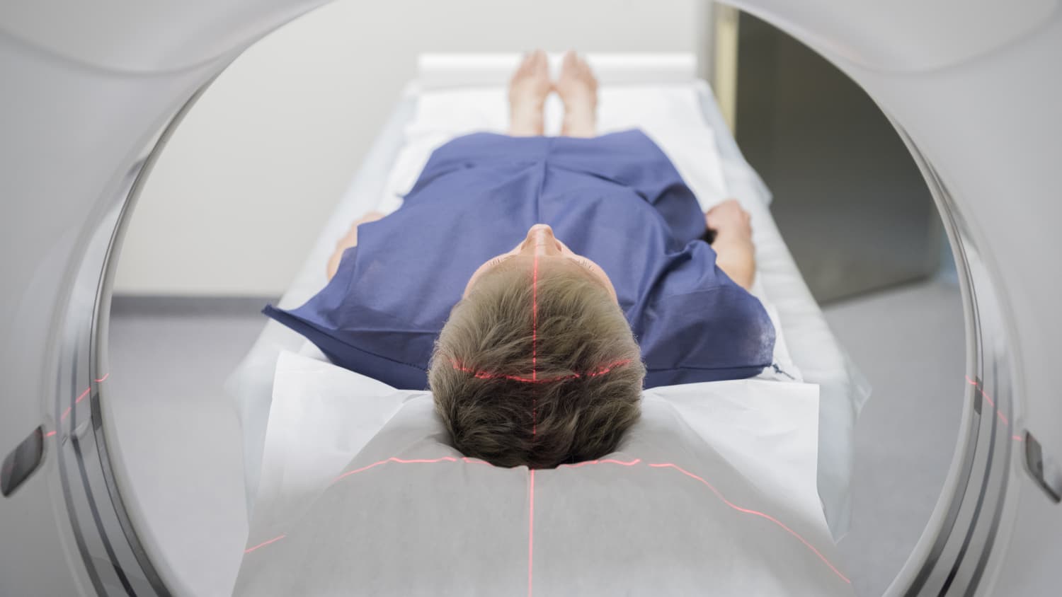
The procedure for a functional MRI is very similar to that of a regular MRI. The patient lies face-up on a flat surface and is rolled into a long tubular machine. The process is painless, although some people may feel claustrophobic or be bothered by the loud noises the machine makes during the scans.
Inside the scanner, patients are given instructions that are displayed digitally inside a pair of goggles—similar to a virtual reality (VR) headset. The tasks are simple, such as squeezing the left hand or thinking of certain words. The functional regions of the brain that light up in the scanner are then combined with regular MRI imaging of the patient’s brain anatomy.
“Once we map out those basic functions, we help surgeons figure out a safe approach for removing a lesion or tumor from the brain—or for surgery for epilepsy patients,” Dr. Zucconi says.
The main difference between the two procedures is that during an fMRI, doctors give the patient instructions and ask him or her to complete silent brain exercises while lying still.
The exercises increase activity in specific parts of the brain, increasing blood flow and oxygen to them. This activity lights up on the images created by the scanner, giving doctors a visible record of an exact map of the patient’s brain.
A normal MRI of the brain can last between 20 to 30 minutes, while the fMRI lasts between 40 to 55 minutes.
What happens after an fMRI?
If a surgery is not scheduled immediately, patients may review the images with their doctors and decide how to proceed. If a tumor partially overlaps with the motor-skills or language center, for example, the patient may choose to only have part of it removed, or to treat the tumor with radiation instead of surgery.
What are the risks and benefits of fMRI?
An fMRI is safe, painless, and noninvasive. There are no known health risks associated with the procedure, as long as the patient has no metal or electronic implants (because the MRI machine has a very powerful magnet).
The benefits, on the other hand, are significant. Before the invention of fMRI, the only way to locate a person’s language or motor-skills center was to stimulate parts of the brain during an operation or perform invasive angiographic examinations—both of which required the patient to be awake to respond to questions. Knowing this information ahead of time makes surgery safer and faster, and the patient can stay under sedation during surgery.
What is unique about Yale’s approach to the fMRI procedure?
Neuroradiologists at Yale Medicine work closely with surgeons, oncologists, and neurologists with specialized knowledge about treating brain tumors and disorders of all types.
“Our ongoing research studies rely on fMRI to better understand various diseases, including psychiatric conditions like depression, as well as Tourette syndrome and epilepsy,” Dr. Zucconi says.
Another area of active research relies on resting state fMRIs. These work as the name suggests, by imaging the brain while it is at rest. “This type of imaging gives insight into the brain’s networks and interconnected regions,” Dr. Zucconi says.