Fetal Ultrasound
Overview
Since the mid-20th century, ultrasound has been used extensively to create a "picture" of what's happening inside our bodies. For pregnant women, it is an indispensable aspect to care.
At Yale Medicine Maternal-Fetal Medicine, our physicians and sonographers perform more than 16,000 ultrasounds a year.
"Experience counts. This is the only way we can approach the fetus like a patient. With sophisticated ultrasound, we now have ways to identify fetal problems and guide treatment," says Joshua Copel, MD, a Yale Medicine expert in prenatal diagnosis and fetal therapy. "If a baby has an abnormal amount of fluid in the chest, we can use an ultrasound-guided needle to drain it out. Or we can deliver blood to the umbilical cord. The first of these types of transfusions were done here at Yale in 1984."
What is ultrasound?
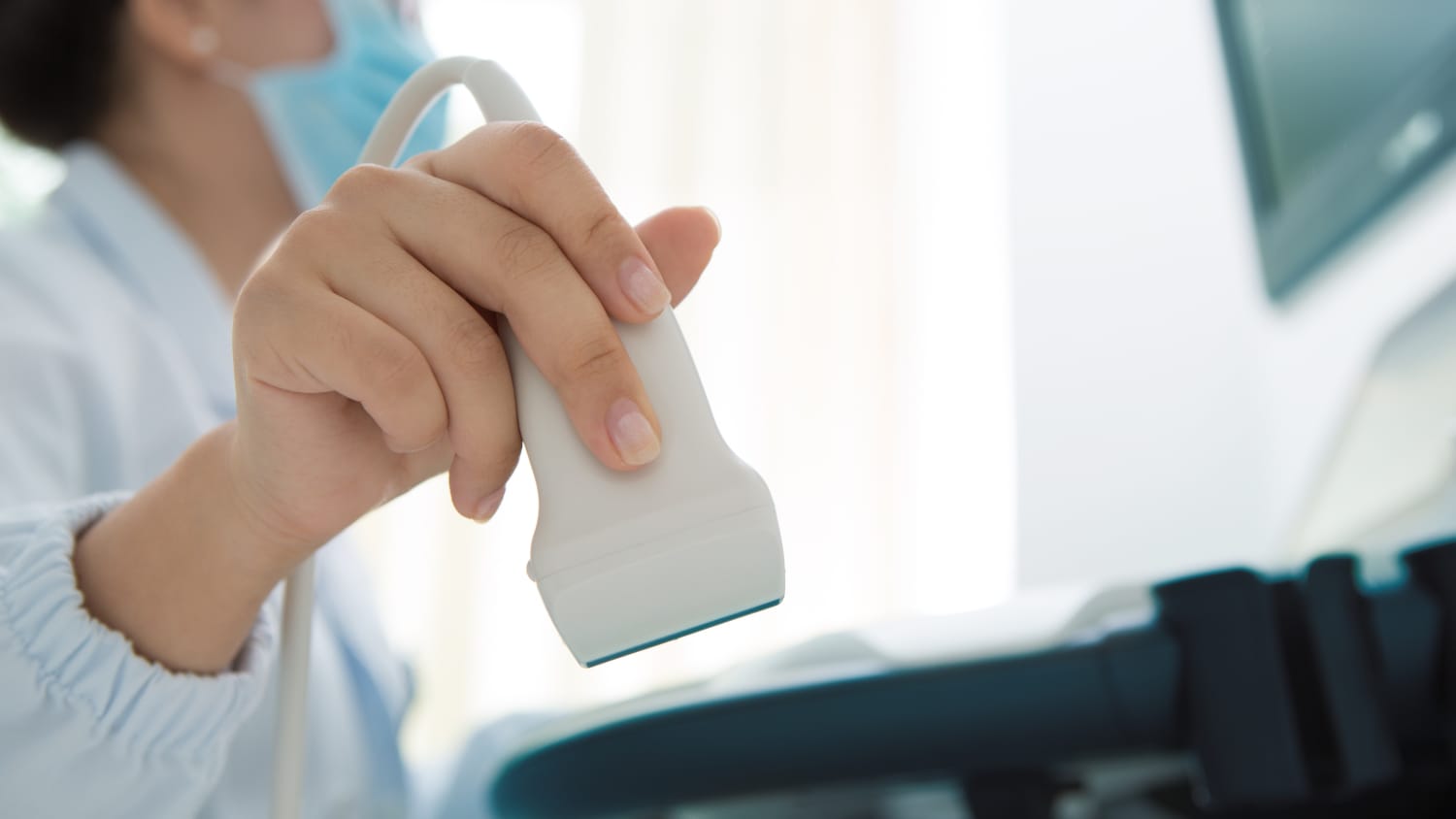
Think of ultrasound—sometimes called a sonogram—as sonar for the body. A small instrument called a “transducer” transmits high-frequency sound waves, bouncing them off structures in the body and using the reflected sound waves to paint a picture.
The sound waves can determine the distance, size, shape and consistency of an object. A computer compiles these results to create an image of the object, be it a fetus, an adult heart, or a tumor in the kidney.
Who performs an ultrasound?
Specially trained physicians and sonographers perform ultrasounds by spreading gel on the skin adjacent to the body part to be imaged. (The gel helps to conduct the sound waves and makes it easier to move the transducer over the skin.) Most ultrasound procedures are painless and non-invasive.
Ultrasound imaging has been used for decades and has an excellent safety record. It’s important to remember that ultrasound uses sound waves; it does not use ionizing radiation, like X-rays, which can cause harm after prolonged exposure
How do ultrasounds assist pregnancy?
Ultrasound is the most widely used medical imaging method for viewing a fetus during pregnancy. It is also utilized to guide procedures such as amniocentesis or chorionic villus sampling.
An ultrasound may be done at almost any time during a pregnancy, even as early as about five weeks after conception. During the first trimester, it can confirm a viable pregnancy and heartbeat, measure the distance from the head to the rear end in order to confirm gestational age, and identify problems such as molar or ectopic pregnancies. It can also identify some uterine or pelvic problems in the mother.
In the second trimester, ultrasound can pinpoint fetal malformations, confirm multiple pregnancies (though this is ideally done in the first trimester), monitor levels of amniotic fluid and evaluate how well the fetus is doing.
In the third trimester, ultrasound can show the placenta's placement, how the fetus is moving and presented for birth, and measure how well the baby is growing.
In what other ways is ultrasound used?
- Abdominal ultrasound can create images of abdominal tissues and organs.
- Ultrasound may guide surgeons as they perform biopsies and other procedures.
- Ultrasound may map brain structures.
- Ultrasound can visualize the interior of the ear or the eye.
- Ultrasound can find clots in veins in the leg.
- It can find bleeding in the abdomen or chest after a motor vehicle accident.
How do you prepare for an ultrasound?
There is no special preparation for a routine ultrasound, though it may make sense to wear loose clothes for comfort. Ultrasounds are painless. Saline used in sonohysterography (a procedure in which saline is injected through the cervix into the uterus, in order to produce a more detailed ultrasound image) occasionally causes mild cramping, but over-the-counter medication is usually enough to manage this pain.
How is Yale Medicine's use of ultrasound technology unique?
At Yale Medicine, we are skilled at using ultrasound to guide care.
"Three percent of babies are born with major birth defects, many of which can be identified with ultrasound," says Dr. Copel. "But it's critical to have experienced eyes and hands do the scans. At Yale, we have both the experience and premium-level equipment with better technology than typically found in other settings."
We have pioneered the use of transvaginal ultrasound early in pregnancy, as well as the use of ultrasound to diagnose pelvic pain, breast cancer and ectopic pregnancy. Yale physicians continue to explore the use of ultrasound to map the brain and track the vascular behavior of cancerous tumors.
We are also exploring the use of Doppler ultrasound to monitor cardiovascular conditions.